|
In a stunning discovery that overturns decades of
textbook teaching, researchers at the University of
Virginia School of Medicine have determined that the
brain is directly connected to the immune system by
vessels previously thought not to exist.
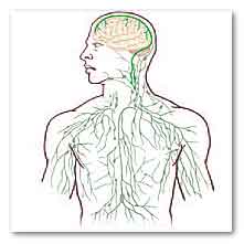
In searching for T-cell gateways into and out of the
meninges, researchers discovered functional
lymphatic vessels lining the dural sinuses. These
structures express all of the molecular hallmarks of
lymphatic endothelial cells, are able to carry both
fluid and immune cells from the cerebrospinal fluid,
and are connected to the deep cervical lymph nodes.
The unique location of these vessels may have
impeded their discovery to date, thereby
contributing to the long-held concept of the absence
of lymphatic vasculature in the central nervous
system.
“Instead of asking, ‘How do we study the immune
response of the brain?,’ ‘Why do multiple sclerosis
patients have the immune attacks?,’ now we can
approach this mechanistically – because the brain is
like every other tissue connected to the peripheral
immune system through meningeal lymphatic vessels,”
said Jonathan Kipnis, a professor in U.Va.’s
Department of Neuroscience and director of U.Va.’s
Center for Brain Immunology and Glia. “It changes
entirely the way we perceive the neuro-immune
interaction. We always perceived it before as
something esoteric that can’t be studied. But now we
can ask mechanistic questions."
He added, “We believe that for every neurological
disease that has an immune component to it, these
vessels may play a major role. [It’s] hard to
imagine that these vessels would not be involved in
a [neurological] disease with an immune component.”
The discovery was made possible by the work of
Antoine Louveau, a postdoctoral fellow in Kipnis’
lab. The vessels were detected after Louveau
developed a method to mount a mouse’s meninges – the
membranes covering the brain – on a single slide so
that they could be examined as a whole. “It was
fairly easy, actually,” he said. “There was one
trick: We fixed the meninges within the skullcap, so
that the tissue is secured in its physiological
condition, and then we dissected it. If we had done
it the other way around, it wouldn’t have worked.”
After noticing vessel-like patterns in the
distribution of immune cells on his slides, he
tested for lymphatic vessels and there they were.
The impossible existed.
As to how the brain’s lymphatic vessels managed to
escape notice all this time, Kipnis described them
as “very well hidden” and noted that they follow a
major blood vessel down into the sinuses, an area
difficult to image. “It’s so close to the blood
vessel, you just miss it,” he said. “If you don’t
know what you’re after, you just miss it.
“Live imaging of these vessels was crucial to
demonstrate their function, and it would not be
possible without collaboration with Tajie Harris,”
Kipnis noted. Harris is an assistant professor of
neuroscience and a member of the Center for Brain
Immunology and Glia. Kipnis also saluted the
“phenomenal” surgical skills of Igor Smirnov, a
research associate in the Kipnis lab whose work was
critical to the imaging success of the study.
The unexpected presence of the lymphatic vessels
raises a tremendous number of questions that now
need answers, both about the workings of the brain
and the diseases that plague it.
For example, take Alzheimer’s disease. “In
Alzheimer’s, there are accumulations of big protein
chunks in the brain,” Kipnis said. “We think they
may be accumulating in the brain because they’re not
being efficiently removed by these vessels.” He
noted that the vessels look different with age, so
the role they play in aging is another avenue to
explore.
And there’s an enormous array of other neurological
diseases, from autism to multiple sclerosis, that
must be reconsidered in light of the presence of
something science insisted did not exist.
The findings have been published online by the
prestigious journal Nature and will appear in a
forthcoming print edition. The article’s authors are
Louveau, Smirnov, Timothy J. Keyes, Jacob D. Eccles,
Sherin J. Rouhani, J. David Peske, Noel C. Derecki,
David Castle, James W. Mandell, Lee, Harris and
Kipnis.
For more information
Nature
Structural and functional features of central
nervous system lymphatic vessels
University of Virginia School of Medicine
MDN |