|
A research, led by team of young researchers at the
Institute of Applied Science and Intelligent Systems
of the National Council of Research in Pozzuoli,
Napoli, in collaboration with the Ceinge-Advanced
Biotechnologies, which involves University Federico
II, reveals an innovative technique for the
identification of 'foreign' and rare cells
circulating in the blood stream, the so called Ctc
(Circulating Tumor Cells).
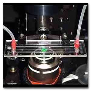
Microfluidic channel used for experiments.
The research has been published in Light: Science
and Applications, a Nature Publishing Group Review.
Blood is composed of millions of cells as red blood
cells, white cells, platelets, lymphoid cells.
At present, the diagnosis of a blood disease is
performed by the blood count, which furnishes
statistical parameters on examined cells as the
cellular volume, haemoglobin, etc.
In order to obtain morphological information,
however, is necessary to study at the microscope the
blood smear, which limits the analysis to a little
fraction of cells and, also, is subjective,
depending on the physician’s interpretation of the
image analyzed.
The results demonstrate that is possible to perform
an in-flow cyto-tomography on liquid samples using a
microfluidic technology, or as it is known
'Lab-on-a-Chip'.
“This new interferometric technique, based on
digital holography, allows us to analyze millions of
cells while flowing in a microfluidic channel,
furnishing parameters as haemoglobin, as same as the
traditional blood count.
Moreover, it is able to analyze every single cell
almost in real time, reconstructing the
three-dimensional image with incredible accuracy”,
illustrate the authors Francesco Merola, Lisa Miccio,
Pasquale Memmolo and Martina Mugnano by Isasi-Cnr.
“ By the very same approach, it would be possible to
identify rare cells, early symptom of potential
pathologies, unseen by a classic analysis. The key
of the technique is to exploit the 360° rotation of
the cells while flowing in the channel, and this
allows us to retrieve the 3D structure of each cell
with dimensions up to one thousandth of millimeter.”
This study has allowed to obtain a tomography of red
blood cells by patients affected with different
types of anemias, identifying them with absolute
accuracy.
“Thanks to the particular sensitivity of this
optical imaging technique, even the smallest
morphological variation compared to the healthy red
blood cell can be revealed, discerning very quickly
and in an objective way the possible related
disease: a sort of liquid biopsy”, says Achille
Iolascon by Ceinge, Full Professor of Medical
Genetics at the Federico II University of Naples.
“Thanks to this technique it will be possible to
study every category of cells, not only the blood
ones”, ends Pietro Ferraro, Director of Isasi-Cnr (www.isasi.cnr.it)
“In fact, thanks also to the contribution of the
colleagues of the Institute of Biomolecular
Chemistry of the Cnr, the validity has been
confirmed also with diatoms - a class of algae
producing more than 20% of the oxygen in the entire
earth - the presence of which in the oceans is a
fundamental signal of the ecosystems health.
Chloroplasts, diatoms’ elements responsible of
photosynthesis, are extremely sensitive to
contaminants in marine water, and this technique
permits to obtain their complete 3D shape, giving
information on a potential contamination”.
The interdisciplinary team of investigators made of
physicists, engineers, biologists and chemists, has
obtained a result that will strongly influence the
oncological diagnosis. This first continuous-flow
complete tomography opens the way to the possibility
of finding the 'needle in a haystack', that is, the
circulating tumor cells, early signal of metastasis
so far elusive.
For more information
Tomographic Flow Cytometry by Digital Holography.
Francesco Merola, Pasquale Memmolo, Lisa Miccio,
Roberto Savoia, Martina Mugnano, Angelo Fontana,
Giuliana D’Ippolito, Angela Sardo, Achille Iolascon,
Antonella Gambale and Pietro Ferraro
Light: Science & Applications (2017) 6, e16241; doi:
10.1038/lsa.2016.241.
Link...
MDN |