|
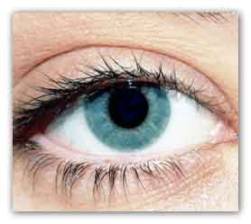
A new study found that neuropsin, a protein in the
retina and cornea, can sense light, but neuropsin is
also expressed in the skin and other parts of the
body. Discovery of that function in cornea raises
hypotheses for undiscovered circadian-rhythm
effects.
Only relatively recently, in 2002, was it proven
that the retina can sense light in its role to help
synchronize our body clocks to Earth’s cycle of
light and darkness.
A just-published discovery – that the cornea is a
light-sensitive tissue, too – has ophthalmologist
Russell Van Gelder excited about the rest of the
body’s potential interplay with circadian rhythms.
“Now we know that people have more photo sensors in
their eye and body than was previously guessed, but
the speculation of what comes next might be the most
exciting aspect of this,” said Van Gelder, director
of UW Medicine’s Eye Institute. He was a co-lead of
the study published recently in PNAS (Proceedings of
the National Academy of Sciences).
The study’s most compelling finding was that
neuropsin, a protein in the retina and cornea whose
function in mammals was heretofore unknown, can
sense light. Retinas and corneas kept in tissue
culture could synchronize their daily rhythms to a
light-dark cycle; retinas and corneas that lacked
neuropsin could not do so.
Neuropsin is also expressed in the skin and other
parts of the body.
“It lets us consider what other types of physiology
might be linked to these photoreceptors, and how we
could co-opt these to help manage diseases,” said
Van Gelder, UW professor and chair of ophthalmology.
“For example, we don’t know exactly what triggers
sun-tanning. That’s an example of a phenomenon that
is light-sensitive but nobody really knows the
receptor for it. We don’t know what causes light
sensitivity in people with lupus and other collagen
vascular diseases, or why light therapy works to
treat certain skin diseases. Your organs may have
access to knowing whether it’s light or dark
outside, and adjust their metabolism appropriately.”
Within the eye, neuropsin now is the sixth working
photopigment scientists have identified. Van Gelder
has long used a camera analogy with patients who
face vision diseases and disorders to explain how
these systems work.
“The cornea and eye’s lens are like the lens of the
camera, focusing light, and the retina is like the
film or the sensor in the back, where the image is
created. For many years people viewed the eye as if
it were an old-style camera, without a light meter.
The discovery of the first non-visual opsin,
melanopsin (1998), identified the first light meter
in the eye. Just like a light meter, melanopsin
measures the brightness of light but it doesn’t
contribute to the image.
“The new opsins, including neuropsin and
encephalopsin, suggest there is not just one light
meter in the eye but multiple light meters that
serve different functions. No one would’ve guessed
that 20 years ago,” he said. “Now our goal is to
figure out exactly how these light meters work and
what functions they control.”
Although this study’s finding spotlighted new
capability of the cornea, Van Gelder said, it also
suggests that the retina is more complex than was
previously suspected.
“We didn’t think the retina needed another
photopigment; it has five we already know about.
What’s remarkable is that it doesn’t use any of
those pigments to synchronize its own circadian
rhythms to the light-dark cycle.”
“Figuring out why evolution found advantage in using
neuropsin is a question that will engage us for the
foreseeable future.”
UW research assistant professor Ethan Buhr was first
author of this work, which was done in collaboration
with the laboratories of King Wai Yau at Johns
Hopkins University and Richard Lang at Cincinnati
Children’s Hospital. The study was supported by
National Institutes of Health grants F32EY02114,
EY14596, EY23179 and EY001370.
For more information
Neuropsin (OPN5)-mediated photoentrainment of local
circadian oscillators in mammalian retina and cornea
link...
MDN |